Mrs. Dewi was 39 years old when she was diagnosed with cavernoma, a blood vessel malformation that causes concealed bleeding in the brain at the end of 2012. This resulted in severe headaches and balance issues. Due to its location in the area between the cerebellum and close to the brainstem, surgery posed a high risk. Mrs. Dewi then chose an alternative treatment using ECCT. Although ECCT was not initially developed for non-cancer cases, it turned out to help improve the blood vessels that were swollen. The dizziness symptoms relatively diminished within a few weeks of using the device. Mrs. Dewi only used the device for a year starting from early 2013 because the headaches had subsided. However, five years later, in 2018, the symptoms reappeared, and she had to use the ECCT device again regularly. It appears that ECCT also helps improve problematic blood vessels like those with cavernoma. However, if the device is stopped for a long period, the symptoms may return.

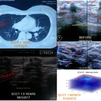
Images: LEFT: Mrs. Dewi’s MRI results when she was first diagnosed with cavernoma in 2012, showing a bleeding lesion in the central cerebellum area approximately 3 cm in size; CENTER: MRI results from 2018 showing the cavernoma lesion reappearing, smaller in size compared to five years earlier after the device was stopped for four years because the symptoms had subsided; RIGHT: MRI results from 2023 showing a small cavernoma lesion still present, but with a solid cyst surrounding it, possibly from dead blood vessel tissue initially suspected to be a cavernoma lesion.
Cavernoma is a type of blood vessel abnormality, not a tumor or growing cells, and can occur in the brain, brainstem, or spine, but it is not a result of metastasis. The repair process that occurs in this blood vessel malformation after using ECCT likely takes place through another mechanism, not due to mitosis disturbance in tumor cells when exposed to the electric field generated by the ECCT device.
Cavernoma is a form of venous malformation due to endothelial dysmorphogenesis of a lesion that may have been present since birth. A cavernoma in the brain is called cerebral cavernous malformation (CCM). Although it is referred to as a hemangioma, cavernous hemangioma is not a tumor because it does not show endothelial hyperplasia. The abnormal tissue causes a slowing of blood flow through the cavities. The blood vessels do not form the necessary connections with surrounding cells without the support structure of smooth muscle tissue, causing leakage into the surrounding tissue. Blood leakage can cause various symptoms such as seizures, chronic headaches, reduced vision, balance issues, and sudden cognitive slowdown.
Initially, Mrs. Dewi only used the device for a year because the severe headaches subsided. However, after the device was stopped for about five years, the severe headaches returned. MRI results also showed the lesion had reappeared, but it was relatively smaller than before. The lesion may have nearly disappeared, but because the device was not used for a long period, the malformation resurfaced. Once again, Mrs. Dewi only used the device for a year because her symptoms began to subside again after resuming regular use.
The mechanism by which ECCT improves malformed blood vessels likely involves stimulating the endothelial cells that form blood vessels, causing mild strain that, when continuously applied, may lead to structural improvement in the cells. This mechanism is often observed in the effect of ECCT on normal tissue or cells, resulting in more cohesive tissue. For surface tissue, ECCT also influences the “cleaning” of waste substances that accumulate on the surface, preventing them from interfering with the physiological function of cells or tissue. Therefore, if ECCT is used routinely over the long term, its effects may become permanent.
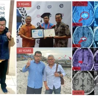
By 2025, Mrs. Dewi will have surpassed 12 years since the initial diagnosis. Her condition remains relatively normal, and she continues to use the device to prevent recurrence. Wishing her continued good health! (WS)