Mrs. Saripah (55 years old) was diagnosed with breast cancer 10 years ago. The biopsy results indicated a malignant invasive ductal carcinoma with moderate differentiation. She chose to rely solely on using the ECCT device, refusing to undergo surgery. An ultrasound after one year of use showed no detectable mass. She continued using the device for five years, during which there was no recurrence and no spread of the cancer. The use of the device was stopped after that since there was no more cancer detected and no spread without any surgery.
However, after stopping the device and following a COVID-19 vaccination, a new lump appeared in her breast near the original tumor site, though of a different type. The new mass was more encapsulated (lobulated). After resuming ECCT treatment, the previously rounded lump began to soften, and the characteristics of the lump appeared benign.

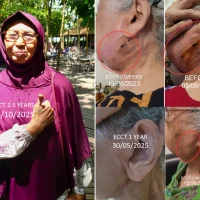
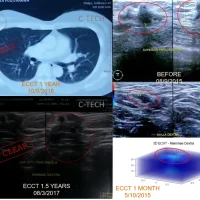
Top right: Mrs. Saripah’s mammography result before using ECCT showed a mass in the left breast’s subareolar area that had already infiltrated the cutis tissue, with microcalcification features. Bottom right: Ultrasound after one year of using ECCT showed the mass was no longer detectable. Top center: ECVT electrical activity scan results showed abnormal activity in the left breast before using ECCT and normalized activity after one year of use. Middle center: Ultrasound showing reappearance of a mass near the original site after 10 years (5 years after stopping ECCT), with benign characteristics. Bottom center: ECVT scans showing recurrent tumor activity in the left breast before resuming ECCT (November 2024) and again after 3 months of use (February 2025), indicating reduced activity close to normal.
After her biopsy, Mrs. Saripah experienced swelling in her armpit and began coughing. She refused a full mastectomy and insisted on continuing ECCT therapy from late 2014. The ECCT device she used was a vest that covered her entire chest, along with a smaller unit targeting the axillary area, worn for 8–12 hours daily.
Dead cells came out through her skin resembling whitish flakes, rich in calcium, likely due to the biopsy having disrupted some of the body’s natural drainage pathways through the blood vessels, urine, and sweat. Her mammogram showed a spiderweb-like (stellate) lesion with heavy calcification—typical of group C2 based on ECCT response classification. This cancer type is usually rich in calcium, spreads easily through the bloodstream to bones and liver, and is often influenced by hormonal activity. C2-type cancers are generally hard to treat with ECCT, are unresponsive to chemo or radiation, and tend to spread aggressively if surgically removed.
The chalk-like skin flakes continued to appear for up to a year. Alongside the discharge, the lump gradually shrank. An ultrasound after one year of ECCT showed no more detectable mass.
Mrs. Saripah continued using the ECCT device for five years, until 2019. Her breast condition had returned to normal, and ultrasound and X-ray results remained consistently normal. However, in 2023, following the pandemic and her COVID-19 vaccination, she felt a new lump in the area of her original biopsy. Ultrasound showed that this mass was different from the original—encapsulated, without calcification, with cystic/necrotic components, resembling an infection. The character matched cancer type A/D based on ECCT response classification.
She resumed ECCT therapy at the end of 2024. The chalk-like flakes began to emerge again from her skin, but this time with less calcium content. The lump gradually began to soften as more flakes were released.
By early 2025, it had been 10 years since Mrs. Saripah’s malignant cancer diagnosis. Alhamdulillah, she is healthy, active, and appears completely normal. May Mrs. Saripah continue to stay healthy (WS).